 Cart(
Cart(


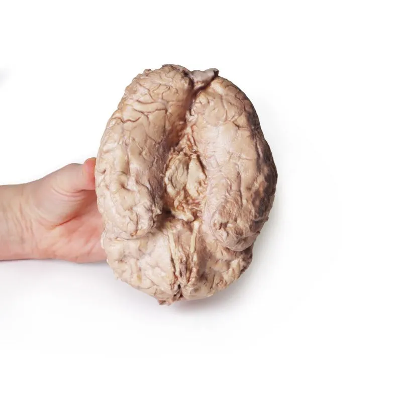

3D Printed Brain (Cerebrum)
This 3D model provides a unique perspective on the anatomy of the cerebrum relative to
the meninges. The cerebrum has been separated from the brainstem and cerebellum, with only parts of the midbrain and
cerebral peduncles visible on the inferior surface. Adjacent to the cut section the olfactory tracts and bulbs can
be seen extending along the inferior margin of the frontal lobes of the cerebrum.
Varying dissection between the left and right cerebral hemispheres allows an appreciation for the organisation of
the brain and meninges as it would normally appear within the cranial cavity. In the midline, the dura mater has
been preserved from anterior (rostral) to posterior. The central portion of the true (endosteal) dura opened to
expose the superior sagittal sinus (between endosteal and meningeal layers of dura mater). Numerous arachnoid
granulations (clusters of arachnoid villi) are visible within the opened superior sagittal sinus – as well as across
the margins of the preserved dura. On the right cerebral hemisphere, the dura mater has been completely removed to
expose the underlying arachnoid mater, which obscures the appearance of the underlying cerebral gyri and sulci as
well as the terminal branches of cerebral arteries. In contrast, the arachnoid mater has dissected across most of
the hemisphere (excepting a margin for reference) to expose the gyri and sulci covered in pia mater. This allows a
clear view of the lateral sulcus and the central sulcus, with the latter defining the boundaries of the frontal and
parietal lobes – and separating the primary sensory and motor cortical areas on the gyri on either side of the
sulcus.
GTSimulators by Global Technologies
Erler Zimmer Authorized Dealer


")


